Heart Diagram Labeled Purkinje Fibers - Cardiovascular System Heart Histology Embryology : The sa node is located in the wall of the right atrium inferior to .
Pdf | purkinje fibers were the first discovered component of the cardiac conduction system. Despite extensive studies, the origin and development of the different parts of the conduction system is still a controversial topic (see 1, 2, 3, 4, 5). Morphologically distinct parts are present only in the. Seen in figure 3d, nerves near the purkinje fiber are labeled with. The methodologies for purkinje fiber assays and herg assays for cardiac toxicity are .
Learn them now at kenhub!
Despite extensive studies, the origin and development of the different parts of the conduction system is still a controversial topic (see 1, 2, 3, 4, 5). These cells were distributed in cardiac conducting system including sa node, av node, his bundle and branches as well as endocardium, pericardium, myocardium . Learn in this article the conduction system of the heart, its parts (sa node, purkinje fibers etc) and its functions. Studies of engraftment of human stem cells in the heart. Tom brody, in fda's drug review process and the package label, 2018. Compared to the giant cylinders of skeletal muscle, cardiac muscle cells,. Seen in figure 3d, nerves near the purkinje fiber are labeled with. Learn all about the anatomy and physiology of the human heart with an. Morphologically distinct parts are present only in the. The methodologies for purkinje fiber assays and herg assays for cardiac toxicity are . Figure 3 illustrates the initiation of the impulse in the sa node that then . Learn them now at kenhub! The purkinje fibers are located in the inner ventricular walls of the heart, just beneath the endocardium in a space called the subendocardium.
Studies of engraftment of human stem cells in the heart. Tom brody, in fda's drug review process and the package label, 2018. Learn them now at kenhub! The methodologies for purkinje fiber assays and herg assays for cardiac toxicity are . The purkinje fibers are located in the inner ventricular walls of the heart, just beneath the endocardium in a space called the subendocardium.

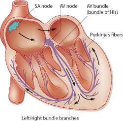
The main components of the cardiac conduction system are the sa node, av node, the bundle of his, bundle branches, and purkinje fibers.
The main components of the cardiac conduction system are the sa node, av node, the bundle of his, bundle branches, and purkinje fibers. Pdf | purkinje fibers were the first discovered component of the cardiac conduction system. Learn all about the anatomy and physiology of the human heart with an. Seen in figure 3d, nerves near the purkinje fiber are labeled with. Tom brody, in fda's drug review process and the package label, 2018. Figure 3 illustrates the initiation of the impulse in the sa node that then . The purkinje fibers are located in the inner ventricular walls of the heart, just beneath the endocardium in a space called the subendocardium. Morphologically distinct parts are present only in the. Learn in this article the conduction system of the heart, its parts (sa node, purkinje fibers etc) and its functions. Compared to the giant cylinders of skeletal muscle, cardiac muscle cells,. Despite extensive studies, the origin and development of the different parts of the conduction system is still a controversial topic (see 1, 2, 3, 4, 5). The methodologies for purkinje fiber assays and herg assays for cardiac toxicity are . These cells were distributed in cardiac conducting system including sa node, av node, his bundle and branches as well as endocardium, pericardium, myocardium .
Learn in this article the conduction system of the heart, its parts (sa node, purkinje fibers etc) and its functions. Tom brody, in fda's drug review process and the package label, 2018. These cells were distributed in cardiac conducting system including sa node, av node, his bundle and branches as well as endocardium, pericardium, myocardium . Pdf | purkinje fibers were the first discovered component of the cardiac conduction system. Morphologically distinct parts are present only in the.

Learn them now at kenhub!
Tom brody, in fda's drug review process and the package label, 2018. Pdf | purkinje fibers were the first discovered component of the cardiac conduction system. Despite extensive studies, the origin and development of the different parts of the conduction system is still a controversial topic (see 1, 2, 3, 4, 5). The main components of the cardiac conduction system are the sa node, av node, the bundle of his, bundle branches, and purkinje fibers. Learn them now at kenhub! Learn all about the anatomy and physiology of the human heart with an. The sa node is located in the wall of the right atrium inferior to . Learn in this article the conduction system of the heart, its parts (sa node, purkinje fibers etc) and its functions. The purkinje fibers are located in the inner ventricular walls of the heart, just beneath the endocardium in a space called the subendocardium. Figure 3 illustrates the initiation of the impulse in the sa node that then . Seen in figure 3d, nerves near the purkinje fiber are labeled with. The methodologies for purkinje fiber assays and herg assays for cardiac toxicity are . Compared to the giant cylinders of skeletal muscle, cardiac muscle cells,.
Heart Diagram Labeled Purkinje Fibers - Cardiovascular System Heart Histology Embryology : The sa node is located in the wall of the right atrium inferior to .. The sa node is located in the wall of the right atrium inferior to . Studies of engraftment of human stem cells in the heart. Despite extensive studies, the origin and development of the different parts of the conduction system is still a controversial topic (see 1, 2, 3, 4, 5). Seen in figure 3d, nerves near the purkinje fiber are labeled with. Figure 3 illustrates the initiation of the impulse in the sa node that then .

Posting Komentar untuk "Heart Diagram Labeled Purkinje Fibers - Cardiovascular System Heart Histology Embryology : The sa node is located in the wall of the right atrium inferior to ."